Fig. 4.
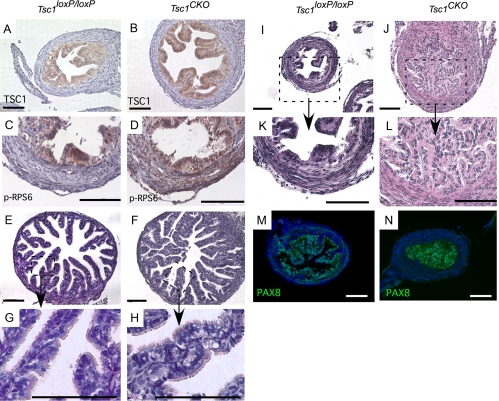
Occlusion of the proximal oviduct in Tsc1CKO mice. A and B, TSC1 expression was detected by IHC in the epithelial cells of both control Tsc1loxP/loxP and Tsc1CKO mice. C and D, IHC for p-RPS6 was used to show evidence for TSC1 deletion in the stroma of Tsc1CKO oviducts. Nuclei are stained with hematoxylin in A–D. E–H, H&E of sections from typical distal portion of oviducts isolated from control and mutant Tsc1CKO mice (E and F) show that the ciliated and secretory epithelial cells of control and mutant distal oviducts are intact (G and H). I–L, Disorganization and dysplasia of secretory epithelial cells of the proximal portion of the oviduct in the mutant females was observed that appeared to occlude the lumen, which might disturb proper transit of embryos. G, H, K, and L are higher magnification images of the boxed areas of E, F, I, and J, respectively. M and N, Immunofluorescence detection of PAX8 in the epithelial cells is shown in the control and the occluded mutant proximal oviducts. Nuclei are stained with 4′,6-diamidino-2-phenylindole. Photos are representative of at least n = 3 for controls and mutants. Bars, 100 μm.

