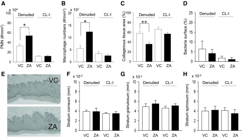
Fig. 5.
Histomorphometric findings in the oral mucosa. Significantly more inflammatory (PMN) cells (A) and macrophages (B) were found in the connective tissues of the denuded side in ZA-treated mice vs. control mice. The numbers of inflammatory cells (A) and macrophages (B) were low and similar between groups in the connective tissues of the contralateral intact side. C, Collagenous tissue areas were assessed in Masson's trichrome-stained sections of the maxillae. The collagenous tissue area in the denuded side was significantly smaller in ZA-treated mice vs. control mice. D, Gram (+) and (−) bacteria were stained, and the bacterial surface per epithelial surface was quantified. No differences were detected between groups. E, Representative photomicrographs of HE-stained regenerated oral mucosa. F and H, The epithelium were histomorphometrically analyzed. No differences were noted in the thickness of stratum corneoum, granulosum, and spinosum between groups. n = 7/group; *, P < 0.05; **, P < 0.01.

