
Figure 5.
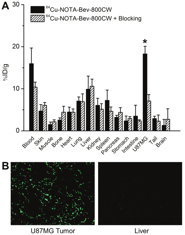
Biodistribution and histology studies. (A) Biodistribution data at 72 h post-injection of 64Cu-NOTA-Bev-800CW, or 2 mg of Bev before 64Cu-NOTA-Bev-800CW (i.e. blocking). *: P < 0.05 (n = 4). (B) Immunofluorescence VEGF staining of the U87MG tumor and liver tissue sections. Bev and AlexaFluor488-labeled goat anti-human IgG were used for VEGF staining. Images were acquired under the same condition and displayed at the same scale. Magnification: 200×.