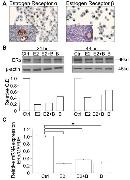
Fig. 4.
K-ras/Pten mouse OvCa cell line hormone receptor characterization.
(A) The K-ras/Pten mouse OvCa cell line expresses ERα and ERβ. Cell blocks were made from the K-ras/Pten mouse OvCa cell line and stained by immunohistochemistry for ERα and ERβ. Inset positive control = mouse uterus (ERα) and mouse ovarian follicle (ERβ). (B) The K-ras/Pten mouse OvCa cell line was treated for 24 or 48 hours with hormones in charcoal stripped phenol red free complete media, followed by western blots for ERα and β-actin. Protein expression was quantitated and normalized to β-actin using NIH Image J software. Columns, fold-change in ERα optical density (OD) compared to placebo. (C) The K-ras/Pten cell line was treated with the indicated hormones for 24 hours then total RNA was extracted and the relative expression of ERα normalized to glyceraldehyde-3-phosphate dehydrogenase was measured using TaqMan quantitative real-time RT-PCR.
Groups: Ctrl= vehicle control, E2= 17β-estradiol, E2+ B= 17β-estradiol plus bazedoxifene, B= bazedoxifene alone

