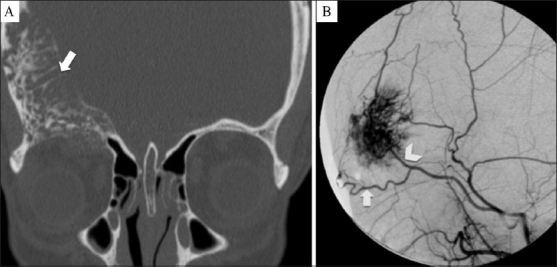
Figures 2 (A, B).

Coronal CT scan (A) shows a large intraosseous mass with prominent vascular channels causing thickening of the frontal bone and right orbit roof (arrow). Digital subtraction angiography (B) shows the intense blush of the mass, fed by the supraorbital (arrow) and the superficial temporal (arrowhead) arteries
