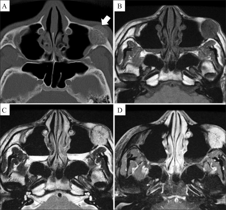
Figures 4 (A-D).

Axial CT scan (A) shows the characteristic radiating appearance of an intraosseous cavernous hemangioma (arrow) arising in the left zygomatic-maxillary fissure. The mass shows low signal (arrow) on the axial T1W MRI image (B), high signal (arrow) on the axial T2W MRI image (C), and avid enhancement (arrow) on a contrast-enhanced T1W fat-saturated MRI image (D)
