Figure 1 (A,B).

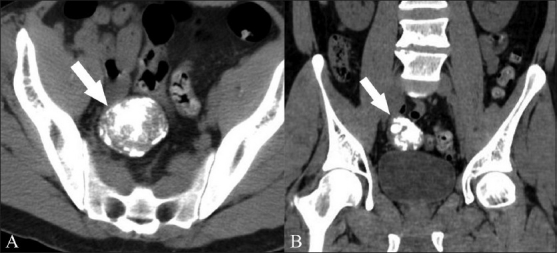
Axial (A) and coronal (B) noncontrast CT scans show a 5 cm, densely calcified pelvic mass (arrow) and its intimate relationship with the adjacent small bowel loops

Axial (A) and coronal (B) noncontrast CT scans show a 5 cm, densely calcified pelvic mass (arrow) and its intimate relationship with the adjacent small bowel loops