
Figure 3.
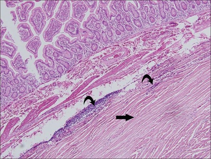
Low-power (10×) photomicrograph stained with hematoxylin and eosin shows relatively hypocellular spindle cell proliferation (bottom right corner), with dense collagen fibers (arrow) and scattered to loosely aggregated lymphocytes and plasma cells (curved arrows)