
Figure 6. IMPs form neurospheres at 37°C in the presence of bFGF.
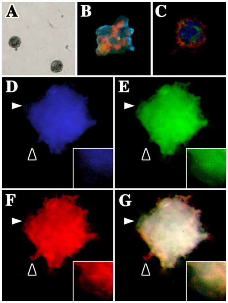
IMPs were cultured at low density in stem cell culture medium containing 20 ng/ml bFGF. (A) IMP-derived neurospheres are formed within two weeks. Spheres were pulse-labeled with 10 μM BrdU overnight and fixed and dual labeled with antibodies against (B) BrdU (green) and nestin (red) or (C) NG2 (green) and PDGFβR (red). Nuclei are labeled with DAPI (blue). (D–G) IMPs differentiate into each of the major cell types in the CNS: oligodendrocytes, astrocytes and neurons. Three-week-old neurospheres were triple-labeled with antibodies against GFAP (blue), O4 antigen (green) and neurofilament (red). (G) The three-channel overlay shows that all antigens generally overlap (white); however, insets in D–F show high magnification images (captured at the white arrowheads) to demonstrate that the staining patterns are not identical. Black arrowheads also highlight non-uniform labeling for the three antibodies.