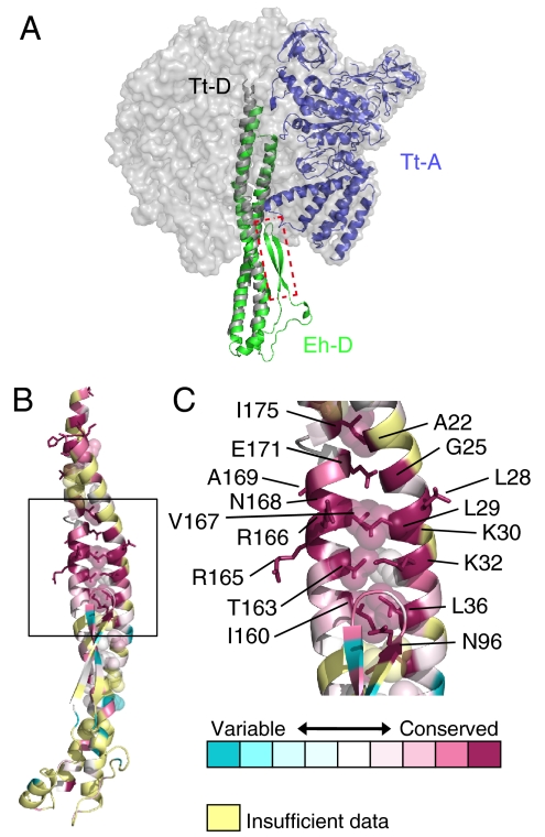
Fig. 3.
Structural similarity of the D subunit. (A) Superimposed structures of Eh-D (green) and Tt-D (gray) of V1-ATPase from T. thermophilus (PDB ID 3A5D). The adjacent Tt-A is shown in blue. The β-hairpin region (residues 90–108) that was deleted in mutation experiments is shown in the red-dotted box. (B and C) Conserved residues of the D subunit. The figures were generated using ConSurf (35). Residues are colored in accordance with conservation among the aligned amino acid sequences of the D subunit from seven species in Fig. S2.

