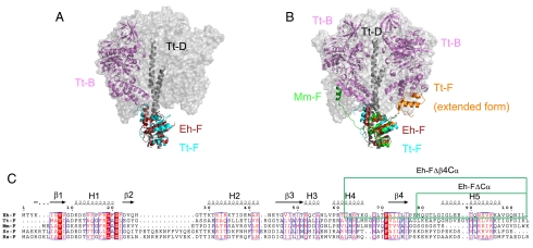
Fig. 4.
Structural similarity of the F subunit. (A) Superimposed structures of Eh-F (dark pink) and the F subunit (cyan) of V1-ATPase from T. thermophilus (PDB ID 3A5D). The adjacent Tt-B is shown in violet. (B) Structures of Tt-F (orange) extended form (PDB ID 2D00) and Mm-F (green) are superimposed in A. (C) The sequence alignment of the F subunits from different species (E. hirae, T. thermophilus HB8, Methanosarcina mazei Gö1, Saccharomyces cerevisiae, and Homo sapiens). The secondary structures of Eh-F are shown above the sequence. The deleted regions of the mutations are indicated in the green box.

