Fig. 5.
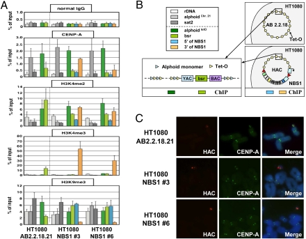
Immuno-FISH and ChIP analyses of the alphoidtetO-HAC/NBS1. (A) ChIP analysis of centromeric chromatin in alphoidtetO-HAC/NBS1 clones 3 and 6. Normal mouse IgG (Top), antibodies against CENP-A, H3K4me2, H3K4me3, and H3K9me3 were used for analysis. The assemblies of these proteins on the alphoid DNA of the original alphoidtetO-HAC in AB2.2.18.21 cell line (Left), the alphoidtetO-HAC carrying the human NBS1 gene in NBS1 no. 3 and NBS1 no. 6 cell lines (Right), are shown. The bars show the percentage recovery of the various target DNA loci by immunoprecipitation with each antibody to input DNA. Error bars indicate SD (n = 2 or 3). Analyzed loci are alphoidtetO (alphoid DNA with tetO motif on the alphoidtetO-HAC), bsr (the marker gene in the HAC vector sequence), and 3′ and 5′ ends of NBS1. rDNA (5S ribosomal DNA), alphoidchr.21 (centromeric alphoid DNA of chromosome 21), and sat2 (pericentromeric satellite 2) were used as controls. Immunoprecipitated DNAs were quantified by real-time PCR. (B) Positions of probes for ChIP analysis in the original HAC and in the HAC carrying the NBS1 gene are shown by colored boxes. (C) Immuno-FISH analysis of metaphase chromosome spreads containing the alphoidtetO-HACs. Cells with the original alphoidtetO-HAC (AB2.2.18.21) and with alphoidtetO-HAC carrying the NBS1 gene (clones 3 and 6) were used for analysis. Immunolocalization of the centromeric protein CENP-A on metaphases was performed by indirect immunofluorescence with anti–CENP-A antibody and Alexa 488-conjugated secondary antibody (green). HAC-specific DNA sequence (BAC) was used as a FISH probe to detect the HAC (red). CENP-A and BAC signals on the HACs overlap one another.

