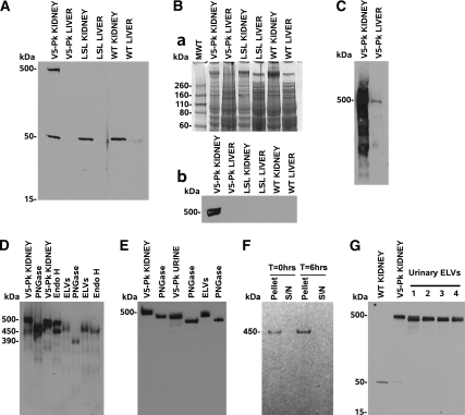
Figure 6.
SV5-Pk tagged fibrocystin is easily detected in the kidney, urine, and urinary ELVs. (A). A) 4–12% MOPS PAGE gel resolving from 15–550 kD, there is a 500 kD band in Pkhd1Pk(+)/Pk(+) kidney membrane preparation but no tagged material was seen in Pkhd1LSL(−)/LSL(−) or WT tissues. Note: there is an endogenous band at 50 kD which cross reacts with SV5–Pk1 antibody (this can be used as a loading control). (B) (a) Coomassie blue staining of a duplicate 4 to 12% MOPS PAGE gel in A; (b) 4% slab gel showing that fibrocystin runs as a doublet. (C) 3–8% Nu–PAGE gel comparing 30 μg of Pkhd1Pk(+)/Pk(+) kidney and liver membrane protein, probed with SV5-Pk1 and a HRP conjugated secondary antibody, the liver contains a small fraction of the kidney fibrocystin as biliary epithelial cells make up about 1% of hepatic cells. (D) 3–8% Nu–PAGE gel. Kidney membrane preparations were treated with PNGase and Endo H. PNGase results in fibrocystin resolving at 450 kD, whereas Endo H causes approximately half the kidney fibrocystin to resolve at 450 kD implying that about half of the fibrocystin is in the ER. In the case of Pkhd1Pk(+)/Pk(+) ELVs, fibrocystin shifts from 450 kD to 390 kD after treatment with PNGase (implying that it has undergone the pro–protein convertase cleavage event upon release from the cell). Upon treatment with Endo H there is a very slight decrease in size, c10 kD, implying that the bulk of the glycosylation is mature complex type carbohydrate (post–Golgi). (E) Distribution of fibrocystin in urine of the Pkhd1Pk(+)/Pk(+) mouse. We can use the SV5–Pk1 antibody to detect fibrocystin in unconcentrated urine and again we detected no smaller forms. (F) 200μl of fresh Pkhd1Pk(+)/Pk(+) mouse urine was harvested and 100μl brought to 1x Complete proteinase. This 100μl was centrifuged at 100,000g for 1 h at 4 °C and the supernatant carefully removed and the pellet resuspended. The remaining 100μl was then incubated at 37 °C for 6 h without proteinase inhibitors and the pellet and supernatant collected. A 3–8% MOPS western showed that all of the fibrocystin remained in the PKD–ELV fraction despite the prolonged incubation at 37 °C, showing that the cleaved fibrocystin is firmly attached to the PKD–ELVs. (G) To exclude the possibility of differential splice forms or other processed forms we ran four different ELV samples on a 4 to 12% gel.

