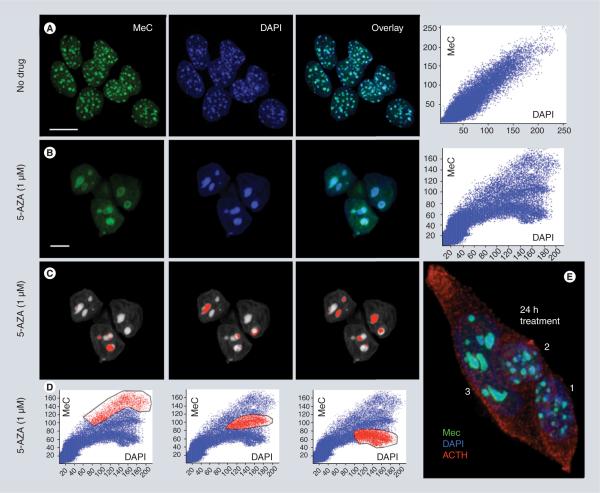
Figure 1. Effect of 5-azacytidine on the heterochromatin of AtT20 pituitary tumor cells in culture.
5-AZA causes both, demethylation of gDNA including DAPI-positive heterochromatic regions and significant reorganization of the chromatin as inferred by maximum intensity projections of three cell nuclei imaged by confocal microscopy, and quantitative 3D image ana lysis (correlated scatterplots). Untreated cells show numerous small (diameter ~1 μm) MeC foci (so-called chromocenters that consist of centromeric and pericentromeric repetitive DNA) – close to the number of different chromosomes – which display an even MeC distribution (in green), and almost fully overlap with the nuclear DAPI signals (in red) (A); in contrast, cells treated for 48 h with 5-AZA show only a few (2–5) giant MeC foci (diameter ~3–5 μm), which mostly exhibit a drastically different MeC distribution. A hypomethylated heterochromatic core surrounded by a hypermethylated ring (B). This phenomenon is represented by changes in the distribution of the MeC and DAPI signals displayed as respective 2D scatter plots (A & B). The reverse mapping of selected groups of plotted signals (red labeled in [D]) explains that corresponding heterochromatic areas of the genome (red labeled in [C]) have experienced different degrees of demethylation in lieu of drug exposure; including loci that stay hypermethylated (left column), loci that have become less demethylated (middle column) and more strongly hypomethylated (right column). Also, drug-treated cells appear larger and flatter than their naive counterparts. (E) The cluster of three ACTH-producing AtT20 cells in this subfigure shows the more heterogeneous drug response of the cells after only 24 h of exposure. The normal-sized nuclei 1 and 2 present foci with unchanged and slightly changed morphology and heterochromatin organization, whereas cell 3 already presents full-blown demethylation effects, including significantly altered morphology and merged chromocenters (scale bars are 10 μm).
5-AZA: 5-azacytidine; ACTH: Adrenocorticotropic hormone; DAPI: 4′,6-diamidino-2-phenylindole; MeC: Methylcytosine.

