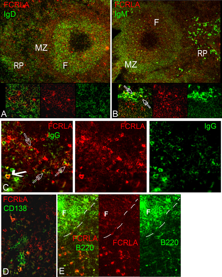
Fig. 5.
Double immunofluorescent staining of spleen cryosections for FCRLA and various lymphocyte markers. (A) IgD/FCRLA: Many of the FCRLAdl cells located in the mantle zone of the follicle coexpress IgD; by contrast, FCRLAbr cells localized outside B-cell follicles are IgD-negative (inset). (B) IgM/FCRLA: FCRLAdl cells of the B-cell follicle coexpress low levels of IgM. Some of the FCRLAbr cells located in the red pulp are strongly stained for IgM (indicated by arrows in inset), while others are weakly stained for IgM or negative. (C) IgG/FCRLA: Some FCRLAbr cells in the red pulp are brightly stained for cytoplasmic IgG (bold arrow), other FCRLAbr cells are weakly stained for IgG (transparent arrows) or negative. (D) CD138/FCRLA: In the red pulp, FCRLAbr cells are found in a close proximity to clusters of CD138-postive plasma cells. Only few of these cells show costaining for both markers. (E) B220/FCRLA: Both FCRLAdl and FCRLAbr cells coexpress B220. However, expression of B220 in T-cell zone FCRLAbr cells is much weaker than that in follicular FCRLAdl cells (the dashed line outlines the border of the follicle). Colors of FCRLA and other markers are as indicated on the figure. F, B-cell follicle; MZ, marginal zone; RP, red pulp.

