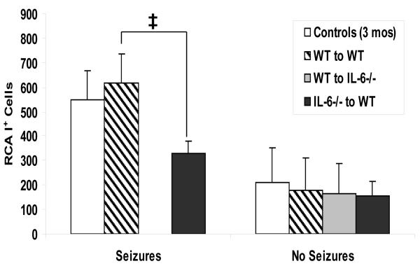
Fig. 2. Activated microglia/macrophages within the hippocampus and dentate gyrus of chimeric mice.
One measure of inflammation is the presence of activated microglia/macrophages, detected through RCA-I lectin histochemistry. The numbers of RCA-I+ cells were enumerated for mice sacrificed on day 14 p.i. There were no irradiated IL-6-deficient chimeric mice reconstituted with IL-6-deficient donor cells (IL-6−/−-to-IL-6−/−) with or without seizures or irradiated IL-6-deficient chimeric mice reconstituted with wild-type donor cells (WT-to-IL-6−/−) with seizures for this time point. The irradiated 3-month-old wild-type chimeric mice reconstituted with wild-type donor cells (WT-to-WT) had a significantly higher number of RCA-I+ cells than the irradiated 3-month-old wild-type chimeric mice reconstituted with IL-6-deficient donor cells (IL-6−/−-to-WT). ‡, p < 0.05 (ANOVA, Fisher’s PLSD post hoc test). Results are mean + SEM of groups with one to fifteen mice per group.

