Figure 1.
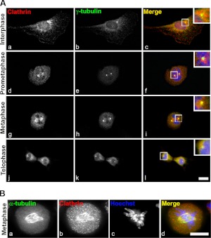
Clathrin is recruited to centrosomes and mitotic spindles during mitosis. A) Analysis of clathrin distribution in cycling NRK cells. Clathrin (red; a, d, g, j) segregates with the γ-tubulin-positive centrosomes (green; b, e, h, k) throughout mitosis. Punctate clathrin staining corresponds to peripheral membrane structures (a, d, g, j). Merged images show colocalization (c, f, i, l); insets show enlarged view of boxed areas. B) Synchronized NRK cells were attached to coverslips by cytospin, probed with antibodies against clathrin (red; a) and α-tubulin (green; b), and DNA or chromosomes stained with Hoechst (i); merged image shows colocalization (d). Images are single sections. Scale bar = 10 μm.

