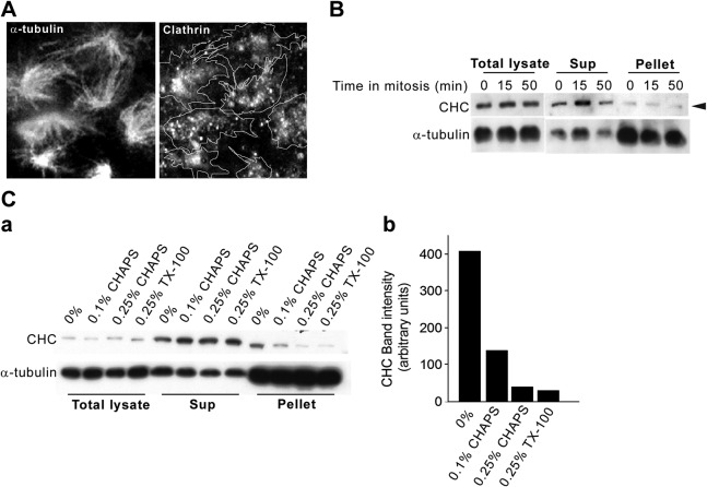
Figure 2.
A large fraction of the spindle-associated clathrin population is membrane bound. A) Mitotic spindles from synchronized NRK cells were prepared in the presence of 0.25% TritonX-100 and were adsorbed onto coverslips and probed with antibodies against α-tubulin and clathrin, followed by immunofluorescence microscopy. Traces in right panel represent outlines of the α-tubulin positive spindles in the left panel. B) Mitotic spindles from synchronized NRK cells were prepared in the presence of 0.25% TritonX-100 and analyzed by Western blotting. Total cell lysates, supernatants (sup), and low-speed pellets (4000 g) from 3 mitotic time points were analyzed (0 min, prometaphase; 15 min, metaphase; 50 min, telophase). C) a) Mitotic spindles were prepared as in panel B, in the absence (0%) or presence of the indicated detergent concentrations (0.1% CHAPS, 0.25% CHAPS, 0.25% TritonX-100) and analyzed by Western blotting with antibodies against CHC and α-tubulin. b) Relative levels of CHC recovered in the α-tubulin pellet in the presence of various detergent concentrations were quantified by densitometric analysis.

