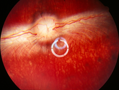
Figure 3.
Rabbit fundus at 5 weeks after high-dose HDP-CDV intravitreal injection. The date show normal retina and clear vitreous without the drug aggregate caused by micelle formulation. The whitish rings in the center were the light reflections from the preposing lens, which renders a wider view of vitreous and retina.

