Fig. 4.
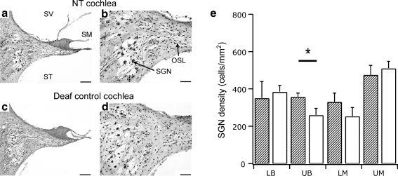
Mid-modiolar sections of the upper basal cochlear region in a deafened cochlea that had received neurotrophins (NTs) without chronic electrical stimulation (a and b) and the deafened untreated contralateral cochlea (c and d). There was a loss of the organ of Corti in both cochleae (a and c) and a small increase in the density of spiral ganglion neurons (SGNs) in the treated cochlea (b) compared to the contralateral cochlea (d). Scale bar = 100 μm (a and c) and 50 μm (b and d). (e) Average SGN density (± SEM) at different cochlear regions in cochleae receiving NTs (diagonal pattern) and the deafened, untreated contralateral cochlea (white bars). There was a significant difference in the mean SGN density (35% greater) restricted to the upper basal region of cochleae receiving NTs compared to the contralateral cochlea (white bars) (paired t test [p < 0.05]). LB = lower basal; LM = lower middle; OSL = osseous spiral lamina; SM = scala media; SV = scala vestibuli; ST = scala tympani; UB = upper basal; UM = upper middle. * - significant difference

