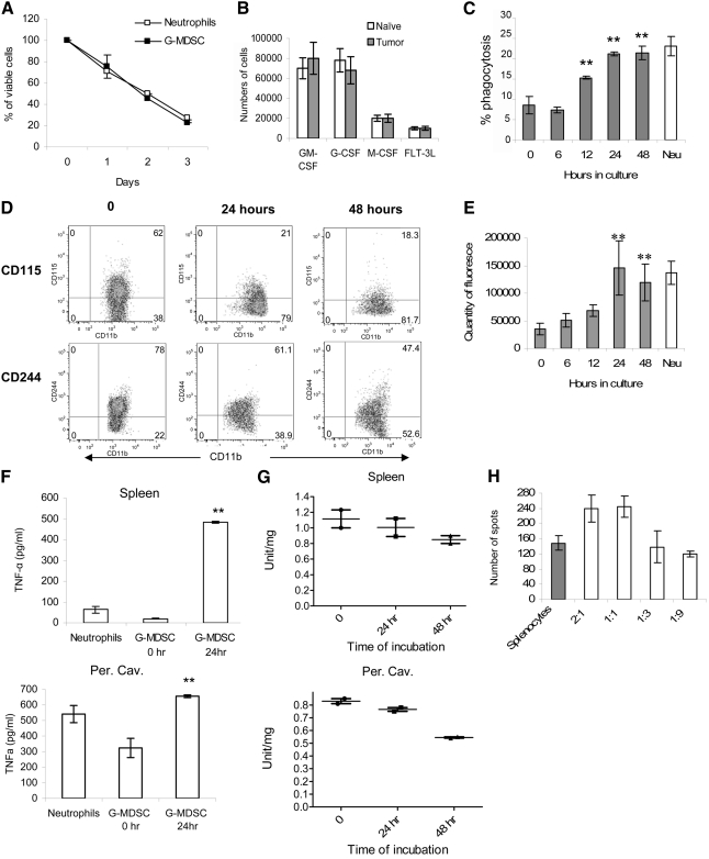
Figure 5. G-MDSC differentiated into Neu in culture.
(A) CD11b+Ly6G+Ly6Chigh cells from bone marrow of naïve or EL-4 tumor-bearing mice were cultured with 10 ng/ml GM-CSF, and their survival was determined at the indicated time. (B) The same cells were cultured for 3 days with 10 ng/ml GM-CSF, 10 ng/ml G-CSF, 10 ng/ml M-CSF, or 100 ng/ml FLT3L. The number of viable cells was assessed by trypan blue exclusion. (C–H) G-MDSCs from spleen of EL-4 tumor-bearing mice were cultured with GM-CSF, and phagocytosis (C), expression of surface markers (D), and LAMP2 (E) were evaluated at the indicated time. Amount of TNF-α (F) and MPO (G) was measured in G-MDSC after cultured with GM-CSF at the indicated time-points. (H) Immune-suppressive activity was measured after 24 h incubation in complete medium. Each experiment was performed at least three times, and cumulative results are shown. **P < 0.01, statistically significant differences from freshly isolated G-MDSC (0 h). Experiments measuring MPO activity were performed twice, and individual results are presented.

