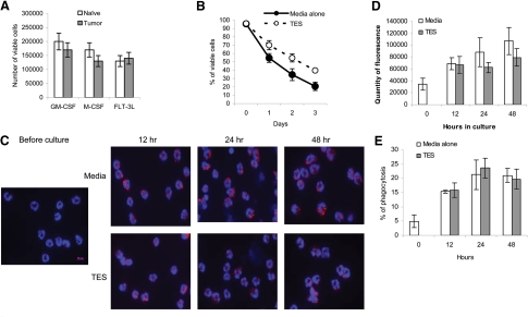
Figure 7. Effect of tumor-derived factors on G-MDSC differentiation.
(A) CD11b+Ly6G+Ly6Chigh cells (2×10⁵) from bone marrow of naïve or EL-4 tumor-bearing mice were cultured with 10 ng/ml GM-CSF, 10 ng/ml M-CSF, or 100 ng/ml FLT3L in the presence of TES for 3 days. Viable cells were assessed using trypan blue. (B–E) G-MDSC from splenocytes of EL-4 tumor-bearing mice were cultured with 10 ng/ml GM-CSF in the presence of TES or control medium, and the viability (B), LAMP2 level (original magnification, ×630; original scale bar=10 μm; C), quantity of fluorescence intensity of LAMP2 (D), and phagocytosis (E) were assessed at indicated time-points. Each experiment was performed at least three times.

