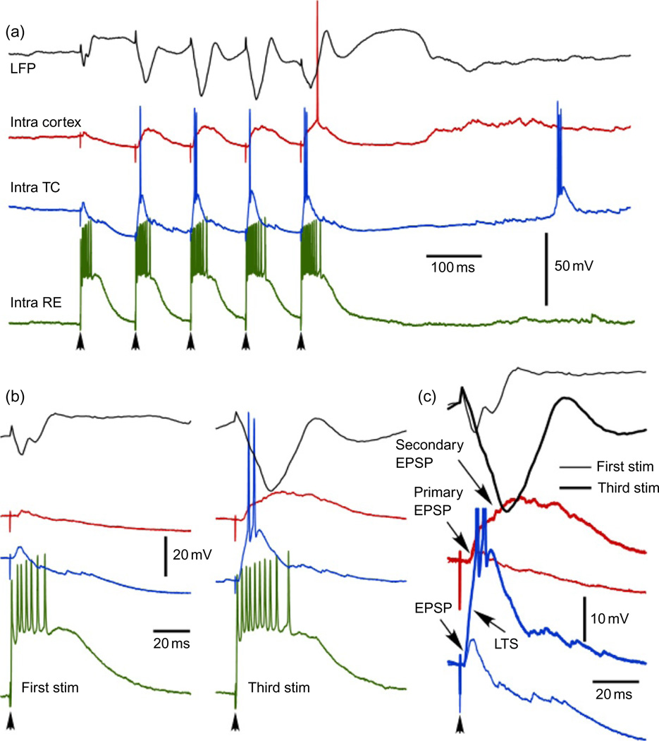
Fig. 3.
Augmenting responses in thalamocortical system. (a) Four different traces show typical responses of motor part of thalamocortical system to 10 Hz pulse train applied to thalamic ventro-lateral (VL) nucleus. Black—local field potential recorded from area 4, red—cortical regular-spiking neurons from area 4, blue—thalmocortical neuron from VL nucleus, green—reticular thalamic neuron from rostrolateral sector of reticular thalamic nucleus. (b and c) Magnified responses to the first and third stimuli. In response to the third stimulus note an increase in the number of spikes in reticular thalamic neuron, generation of LTS in thalamocortical neuron, generation of secondary depolarization in cortical neurons, and a dramatic increase in secondary component of cortical-evoked potential (I. Timofeev, unpublished observations).

