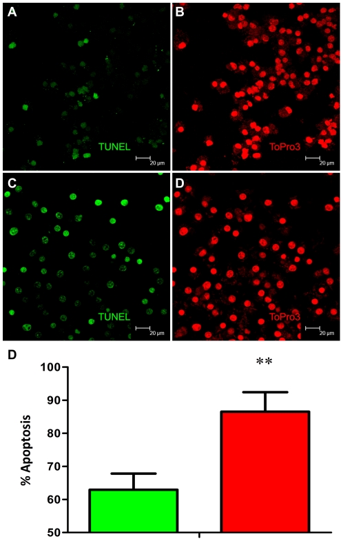
Figure 7. Functional validation of differential expression of apoptotic genes in Rh-BMDM infected with Mtb or Mtb: Δ-sigH by TUNEL assay.
Apoptotic macrophages show TUNEL-positive nuclei stained green . Representative results are shown for cells infected with Mtb (A) and the mutant (C) at the 24-hr post-infection time-point The total number of cells (stained with TOPRO-3 for nuclear DNA in red) can be seen for Mtb (B) as well as the mutant (D). Magnification is to a bar scale. Percentage of macrophage undergoing apoptosis after 24 hours of exposure to Mtb or Mtb: Δ-sigH as quantified by in situ TUNEL assays (E).

