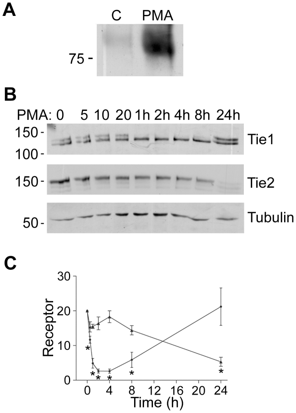
Figure 1. Kinetics of Tie1 and Tie2 Cleavage.
A Tie2 extracellular domain is released from endothelial cells. HUVEC were untreated or stimulated with 10 ng/ml PMA for 24 h, as indicated, before collection of culture medium, removal of cellular material by centrifugation and immunoprecipitation of Tie2 extracellular domain. Immunoprecipitates were resolved by SDS/PAGE and released Tie2 extracellular domain detected by immunoblotting. The position of a 75 kDa molecular mass marker is indicated. B Time course of the effects of PMA on cellular full-length Tie1 and Tie2. Endothelial cells were incubated with 10 ng/ml PMA for the times indicated in minutes or hours (h) before cell lysis and detection of cellular Tie1 by immunoblotting. To determine levels of full-length Tie2 in the same cell population, blots were stripped and re-probed for Tie2. Blots were also re-probed for tubulin. The relative mobility of mass markers is shown in kDa. C Full-length Tie1 (black circle) and Tie2 (black triangle) in endothelial cells treated with 10 ng/ml PMA for different times were determined by immunoblotting in three independent experiments and quantitated by densitometric scanning. Data are means and SEM and are presented as arbitrary units normalized to the levels in untreated cells within each experiment. The asterisk indicates a statistically significant effect of PMA (P<0.05, Students ‘t’ test).

