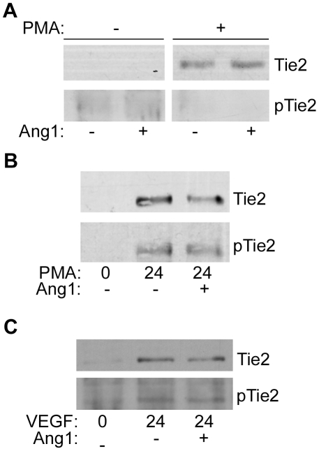
Figure 5. Phosphorylation of Tie2 Endodomain.
A Endothelial cells were incubated for 24 h in the absence of presence of PMA, as indicated, before stimulation with Ang1 for 30 min. Cell lysates were resolved by SDS/PAGE, transferred to nitrocellulose and probed for phospho-Tie2 (pTie2) and Tie2 as indicated. B Following stimulation with PMA for 24 h endothelial cells were incubated with or without Ang1 for 30 min, as indicated. Cells were lysed and Tie2 endodomain recovered from lysates by immunoprecipitation with an antibody recognizing Tie2 intracellular domain and phosphorylation of Tie2 endodomain determined by anti-phosphotyrosine immunoblotting. Blots were stripped and re-probed for Tie2 endodomain. C HUVEC were treated with VEGF for 24 h, with or without Ang1 for 30 min, as indicated. Cells were lysed and Tie2 endodomain recovered from lysates by immunoprecipitation with an antibody recognizing Tie2 intracellular domain and phosphorylation of Tie2 endodomain determined by anti-phosphotyrosine immunoblotting. Blots were stripped and re-probed for Tie2 endodomain. The relative mobility of mass markers is shown in kDa.

