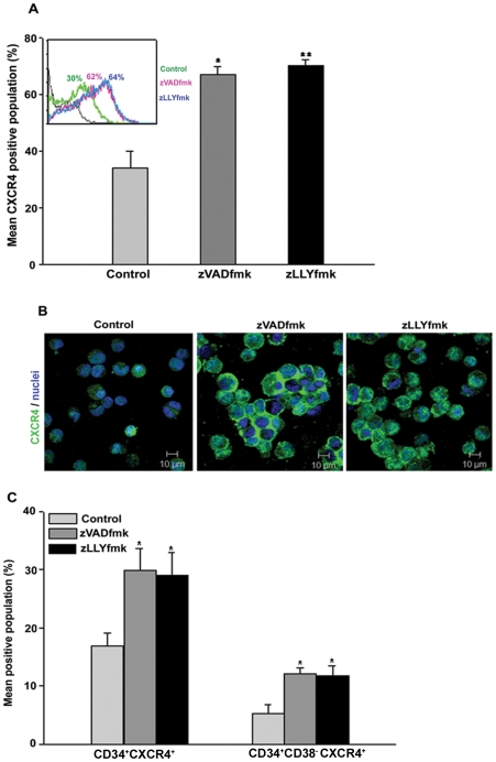
Figure 1. CXCR4 expression analysis of the cultured HSPCs.
(A) The expanded cells were stained using anti CXCR4 antibody and the expression was assessed by flow cytometry. HSPCs cultured in the presence of zVADfmk/zLLYfmk showed a two-fold increase in the CXCR4+ population compared to the control counterpart. Data are represented as mean percentage ± standard deviation of five biological replicates, *p≤0.05, **p≤0.01. A representative flow-overlay is shown in the inset. (B) Confocal microscopy images further confirm higher expression of CXCR4 (green) on cell surface of HSPCs from zVADfmk/zLLYfmk cultures than that of controls. Cell nuclei (blue) were stained with DAPI (bar = 10 µm, n = 3). (C) Flow cytometry analyses of HSPCs after multicolor staining (CD34 and CXCR4 or CD34, CD38 and CXCR4) demonstrate higher CXCR4 positive population in CD34+ and CD34+CD38− compartments. Thus, zVADfmk/zLLYfmk facilitates higher production of CXCR4 expressing CD34+ and CD34+CD38− hematopoietic progenitor cells. Data are represented as mean percentage ± standard deviation of four experiments, *p≤0.5.

