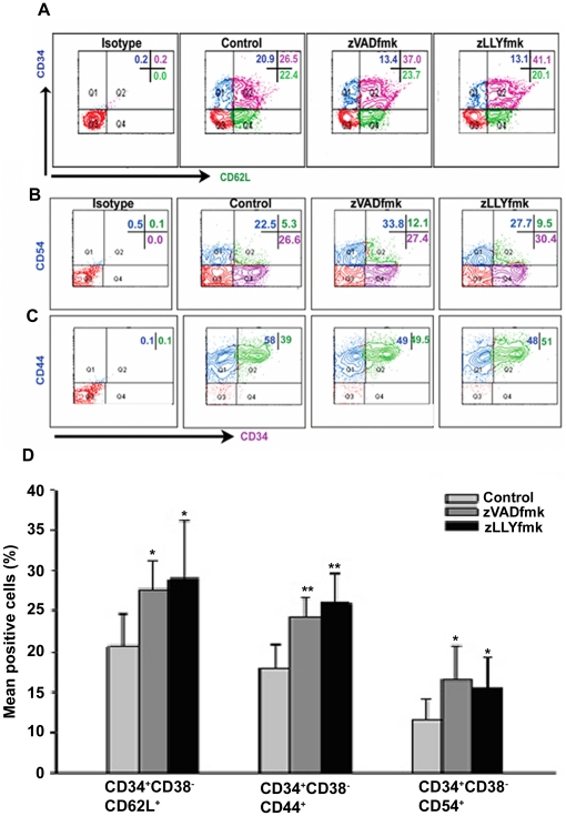
Figure 3. Higher expression of adhesion molecules in the expanded HSPCs.
(A) & (B & C) Flow cytometry analyses of HSPCs after staining for CD34 vs CD62L, CD34 vs CD54 and CD34 vs CD44 antibodies demonstrate higher positive population of these adhesion molecules on CD34+ progenitor cells. (D) Alternatively multi colour flow cytometry analyses showed that zVADfmk/zLLYfmk enhanced the presence of CD62L, CD44 and CD54 on CD34+CD38− subsets as well. Data are represented as mean percentage ± standard deviation of four experiments, *p<0.05, **p<0.01.

