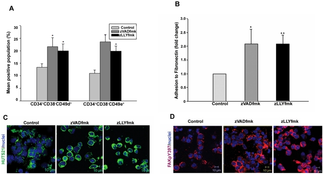
Figure 4. Improved expression of CD49d and CD49e integrins and higher adhesion of the expanded HSPCS.
(A) Multicolour flow cytometry analysis showed a an increase of CD49d and Cd49e integrins in the primitive CD34+CD38− fraction of inhibitor-treated HSPCs. Data are represented as mean percentage ± standard deviation of four experiments, *p<0.05. (B) Adhesion to extracellular matrix protein fibronectin was significantly increased (up to two fold) in the zVADfmk/zLLyfmk cultures compared to the control. Data are represented as mean ± standard deviation of four experiments, *p<0.05. (C) The adhered zVADfmk/zLLYfmk HSPCs retained a higher β1 integrin ligand binding status and the activation of focal adhesion kinase (D) compared to the control, green- HUTS 21, red – FAKpY397, blue – nuclei, scale 10 µm.

