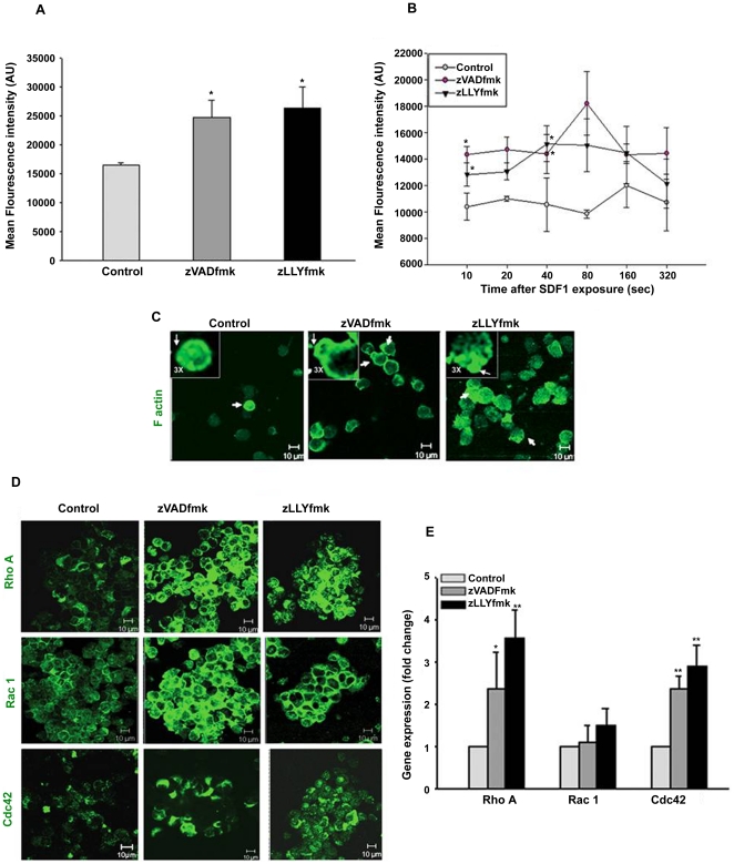
Figure 6. Actin dynamics of expanded HSPCs.
(A) The expanded control and zVADfmk/zLLYfmk HSPCs were assessed for the polymerized actin (F actin) using fluorescein conjugated phalloidin. The presence of zVADfmk and zLLYfmk enhanced the of F actin fluorescence intensity compared to the control. Data are represented as mean ± standard deviation of four experiments, *p≤0.05. (B) Actin polymerization assay done by exposing the control and test HSPCs to SDF1α for different time intervals showed an early response and higher polymerization of actin at all time points analyzed in the zVADfmk/zLLYfmk sets. Data are represented as mean ± standard deviation of three experiments. Statistical analysis was always made between control vs. zVADfmk/zLLYfmk at all time points. *p≤0.05, **p<0.01. (C) The migrated population from the zVADfmk/zLLYfmk HSPCs showed a typical polarized morphology with a prominent localization of F actin towards the leading edge of the cells (white arrow heads & insets) where as in control cells a homogeneous distribution of F actin was seen without much visible cell polarization, green – F actin, bar = 10 µm. (D) The expression of RhoGTPase members RhoA, Rac1 and Cdc42 was found to be increased in the zVADfmk/zLLYfmk HSPCs, green –RhoA, Rac1 and Cdc42, bar = 10 µm (E) Quantitative real time PCR analysis showing a significant up regulation in the gene expression of RhoA upto (3.2 fold) and Cdc42 (up to 2.5 fold) in the zVADfmk/zLLYfmk expanded HSPCs compared to the control, whereas the Rac1 showed a modest increase in those sets. Data are represented as mean ± standard deviation of three experiments, *p≤0.05, **p<0.01.

