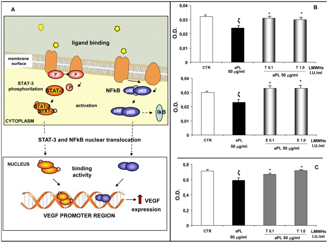
Figure 3. Intracellular mechanisms regulating VEGF expression in HEEC. Effects of aPL and LMWHs on NF-κB and STAT-3 activation.
A. VEGF is a well-known factor able to promote endothelial cell proliferation and new vessel formation. Several intracellular mechanisms regulate VEGF expression in HEEC including NF-kB and STAT-3. The figure illustrates these two signalling pathways whose activation increases VEGF expression. NF-κB activity is regulated by the interaction with the inhibitory IκB protein. Upon activation, IkB is phosphorylated and degraded, thus allowing NF-κB to translocate into the nucleus and bind to VEGF promoter region, upregulating the expression of this proangiogenic factor by HEEC. Moreover STAT-3 is a member of JAK-STAT signalling pathway. It is a latent transcription factor which is activated by phosphorylation. Activated STAT-3 protein induces its nuclear translocation and favours VEGF expression. B. NF-κB activation in the presence of aPL alone or with tinzaparin or enoxaparin after 4 hrs of treatment in differentiation culture medium. aPL reduced NF-κB activation while tinzaparin or enoxaparin were able to restore the NF-κB DNA binding activity. The values are O.D. mean ± SE of three different experiments. CTR: untreated cells; T: tinzaparin; E: enoxaparin; O.D.: Optical Density; ζ: P<0.01 compared with CTR; *: P<0.001 compared with aPL-treatment. C. Effects of aPL on STAT-3 phosphorylation in the presence of tinzaparin after 4 hrs of treatment in differentiation culture medium. aPL reduced STAT-3 activation while tinzaparin was able to restore the STAT-3 phosphorylation. Values are O.D. means ± SE of three different experiments. CTR: untreated cells; T: tinzaparin; O.D.: Optical Density; ζ: P<0.05 compared with CTR; *: P<0.05 compared with aPL treatment.

