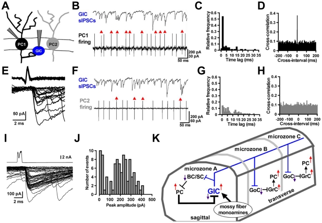
Figure 6. Inhibitory synaptic connections between Purkinje cells and globular cells, and a globular cell-incorporated microcircuit.
(A) Arrangement for paired recordings from a globular cell (GlC) and a Purkinje cell (PC) (PC1 or PC2). (B) Simple spike discharges of PC1 (lower) and sIPSCs in the GlC (upper). Action potentials indicated by red arrowheads caused IPSCs within a 2-ms delay following each action potential. (C) Distributions of time lags between peaks of action potentials in PC1 and onset of IPSCs in the GlC of (B). (D) Cross-correlogram of times of spike-peak and of sIPSC-onset recorded from the same pair in (B). (E) Superimposed traces of the action potentials in the presynaptic PC1 (upper) and individual IPSCs in the GlC (lower). Twenty traces were aligned with respect to the time course of the onset of the presynaptic action potentials. Six spikes failed to evoke IPSCs. (F) A few action potentials of PC2 (lower) caused sIPSCs in the GlC (upper), as shown by red arrowheads. (G) Distributions of time lags obtained in paired recordings from PC2 and the GlC of (F). (H) Cross-correlogram of times obtained from paired recordings in (F). (I) Paired whole-cell recordings from a PC and a GlC. Depolarizing stimulation (−65 to +10 mV, 1-ms duration) was applied to the presynaptic PC with a 2-sec interval. Superimposed fifty traces of presynaptic whole cell currents in the PC (upper) and IPSCs in the GlC (lower) are shown, respectively. (J) The amplitude histogram was obtained from 100 evoked IPSCs recorded in the same pair of (I). (K) A schematic representation of the microcircuit with GlC predicted by the previous anatomical [6] and present studies: the PC-GlC functional connection (thick line) is indicated in the local circuit of cerebellar cortex. BC: basket cell, SC: stellate cell, GoC: Golgi cell, GrC: granule cell.

