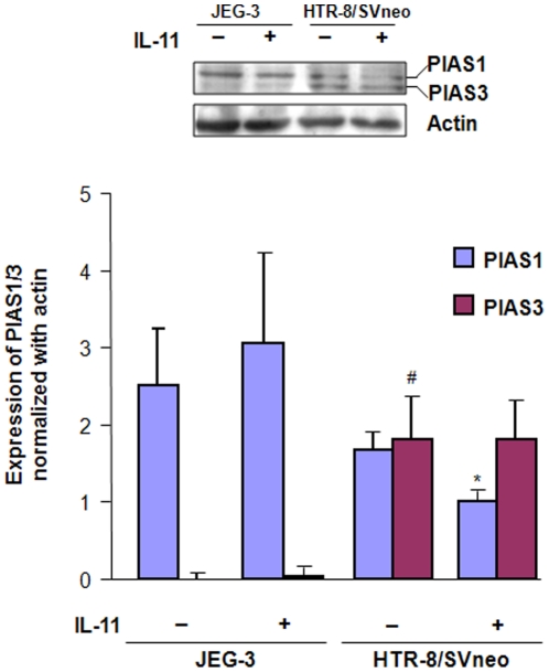
Figure 6. Expression of PIAS1/3 in JEG-3 and HTR-8/SVneo cells following IL-11 treatment.
Cell lysates were prepared after treatment of JEG-3 and HTR-8/SVneo cells with IL-11 (200 ng/ml) for 24 h and Western blot was done for the expression of PIAS1/3 as mentioned in Materials and Methods . Band intensities were normalized with respect to actin and data is expressed as mean fold change in the expression ± SEM of PIAS1 and PIAS3 as compared to the JEG-3 control. *p<0.05 between untreated and IL-11 treated HTR-8/SVneo cells; #p<0.001 between untreated JEG-3 and HTR-8/SVneo cells.

