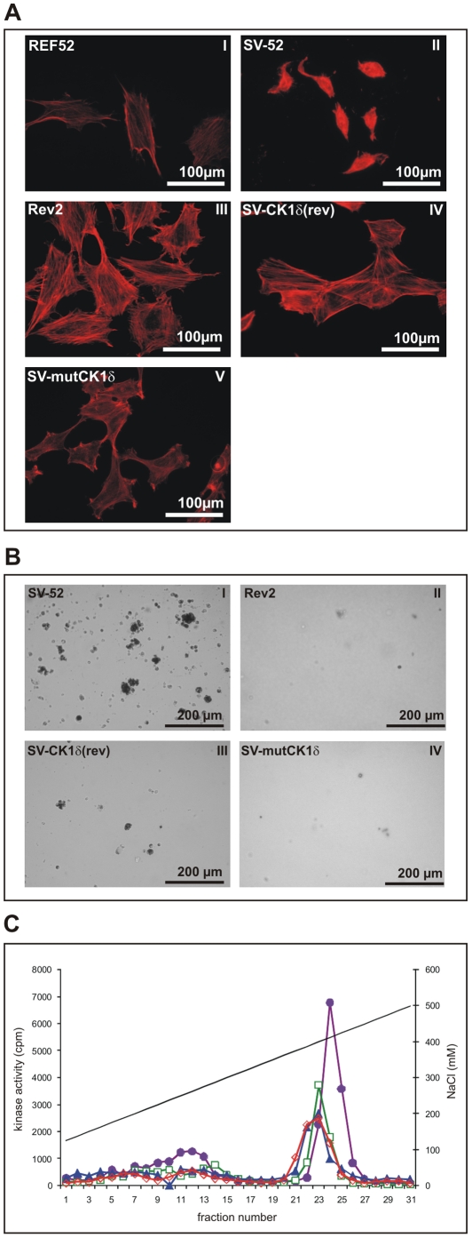
Figure 3. Effects of ectopic expression of CK1δ(rev) or mutCK1δ in SV-52 cells.
(A) Actin network of REF52, SV-52, Rev2, SV-CK1δ(rev) and SV-mutCKδ cells. The actin cable network of parental REF-52 cells (I), maximal transformed SV-52 cells (II), minimal transformed Rev2 cells (III), SV-CK1δ(rev) and SV-mutCKδ cells was stained with phalloidin-TRITC. (B) Colony formation of SV-52, Rev2, SV-CK1δ(rev) and SV-CKmutCK1δ cells in soft agar. Cells were plated in duplicate in culture. Colonies were scored and photographed 20 days after plating (see also table 2). (C) Detection of CK1 activity in cellular protein fractions derived from anion exchange chromatography. Soluble extracts of SV-52 (purple, closed circles), Rev2 (blue, closed triangles), SV-CK1δ(rev) (green, open rectangles) and SV-mutCKδ (red, open rectangles) cells were prepared and in each case equal protein amounts were loaded onto a 1 ml Resource Q column. Then proteins were eluted with a linear gradient of increasing NaCl concentration, 0.25 ml fractions were collected, and kinase activity was determined as described in Materials and Methods. The kinase activities in the peak fractions of SV-52, Rev2, SV-CK1δ(rev) and SV-mutCKδ cells were compared.

