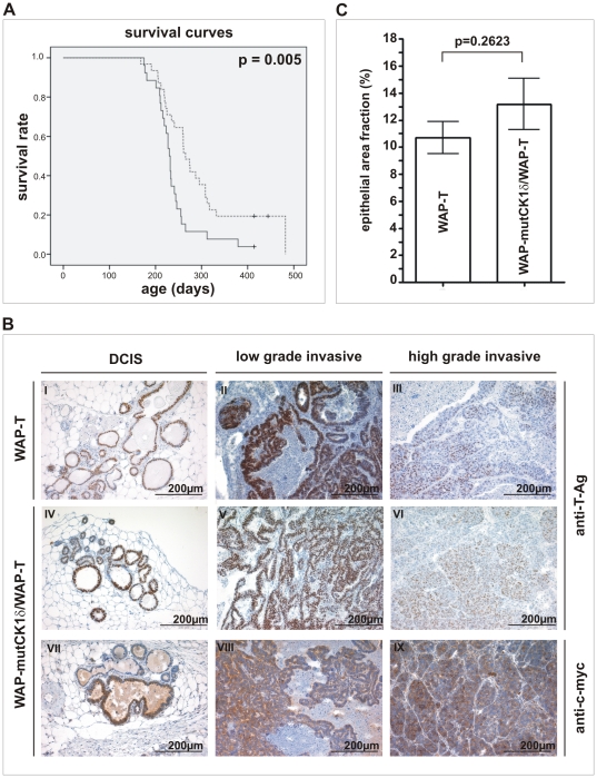
Figure 5. Phenotypic characterization of WAP-mutCK1δ/WAP-T mice.
(A) Survival of induced WAP-T and WAP-mutCK1δ/WAP-T mice. Kaplan-Meier survival curves show a significant longer life-span of WAP-mutCK1δ/WAP-T mice (median: 265 days) compared to WAP-T mice (median: 230 days). p = 0.005; (monoparous females). <$>\raster="rg1"<$> WAP-T mice; <$>\raster="rg2"<$> WAP-T mice censored, --- WAP-mutCK1δ/WAP-T mice; <$>\raster="rg3"<$> WAP-mutCK1δ/WAP-T mice censored. (B) T-Ag and mutCK1δ immunostaining of mammary carcinoma in WAP-T transgenic and WAP-mutCK1δ/WAP-T bi-transgenic mice. Cross-sections of neoplastic mammary glands were immunostained with a polyclonal rabbit antibody against T-Ag (I–VI) and a polyclonal goat antibody against the c-myc epitope tag (VII–IX). Strong nuclear T-Ag staining was detected in DCIS and low grade tumors of WAP-T and WAP-mutCK1δ/WAP-T mice (I, II, IV and V). In high grade tumors, only weak T-Ag expression was found (III, VI). Expression of mutCK1δ, detected by c-myc immunostaining could be found in the cytoplasm and perinuclear region of DCIS (VII) and invasive carcinomas (VIII, IX) of WAP-mutCK1δ/WAP-T mice. (C) Semi-quantitative evaluation of tumor grading in WAP-mutCK1δ/WAP-T mice. Epithelial tissue areas of non-invasive carcinomas in mammary glands from day 60 after induction from both transgenic lines were counted as described in Materials and Methods. No significant difference in the number of non-invasive carcinomas in fractions of epithelial areas could be detected.

