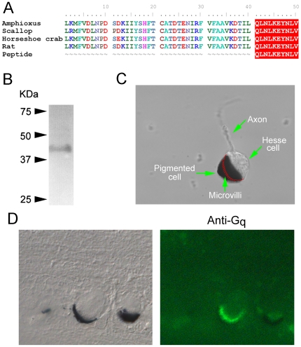
Figure 2. Gq expresses in the microvillar membrane of Hesse cells.
(A) The sequence of the immunogenic peptide used to raise anti-Gq antibodies was aligned with the predicted C-terminal region of Gq of amphioxus, and those of other organisms. (B) Western blot of neural tube using anti Gq, showing the detection of a single band of ≈42 kDa. (C) Morphological characteristics of the Hesse cell: the accessory pigmented cell engulfs the microvilli-covered region of the clear sensory cell; the position of the villous membrane within the occluded region is drawn in red. (D) Left: Nomarski micrograph of a 10 µm section of fixed neural tube containing two Hesse cells. Right: fluorescence image of the same section incubated with anti-Gq antibodies and Alexa Fluo 488-conjugated secondary antibodies. The immunostaining is confined to the region of the microvilli.

