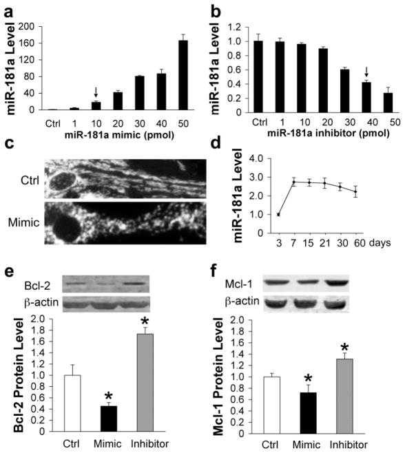
Fig. 4.
Expression of miR-181 and Bcl-2 family proteins after miR-181a mimic or inhibitor transfection of astrocytes. Dose-response of miR-181a levels to transfection with increasing amounts of miR-181a mimic (a) or inhibitor (b) in primary cultures of astrocytes. (c) Mitochondrial morphology changes from a threadlike network (upper) to fragmented round dots (lower) after transfection with 50 pmol miR-181a mimic. Micrographs were taken after staining cells with tetramethylrhodamine methyl ester. (d) Relative miR-181a levels in astrocytes after different durations in vitro. Bcl-2 (e) and Mcl-1 (f) protein expression in primary astrocyte cultures is significantly decreased by transfection with miR-181a mimic and significantly increased by transfection with inhibitor, N=3. All experiments were performed 3 times in triplicate. *P<0.01 compared to Ctrl.

