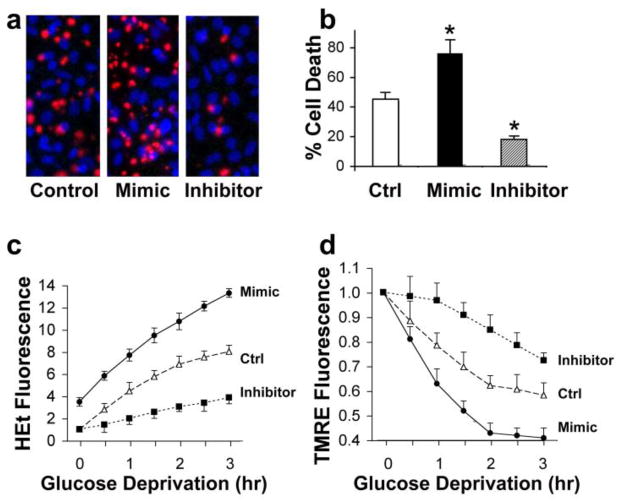
Fig. 5.
Effect of miR-181a mimic and inhibitor on astrocyte ischemia-like injury in vitro. (a) miR-181a mimic aggravates and miR-181a inhibitor reduces injury induced by 24 hr glucose deprivation (GD) in astrocytes. Representative micrographs of cultures stained with propidium iodide (red, dead cells) and Hoechst dye (blue, live cells), are shown. (b) Bar graph shows quantitation of cell death by cell counting. * indicates significantly different compared to control (Ctrl) cultures subjected to the same injury. (c) Transfection with miR-181a mimic or inhibitor affects the time course of reactive oxygen species (ROS) generation. Increasing ROS is detected as increasing HEt fluorescence due to GD stress. (d) Increased miR-181a mimic and inhibitor alter the time course of change in mitochondrial membrane potential induced by GD as assessed by TMRE fluorescence. Mitochondrial depolarization is indicated by decreased fluorescence. All experiments were performed 3 times in triplicate. *P<0.01

