
Figure 5.
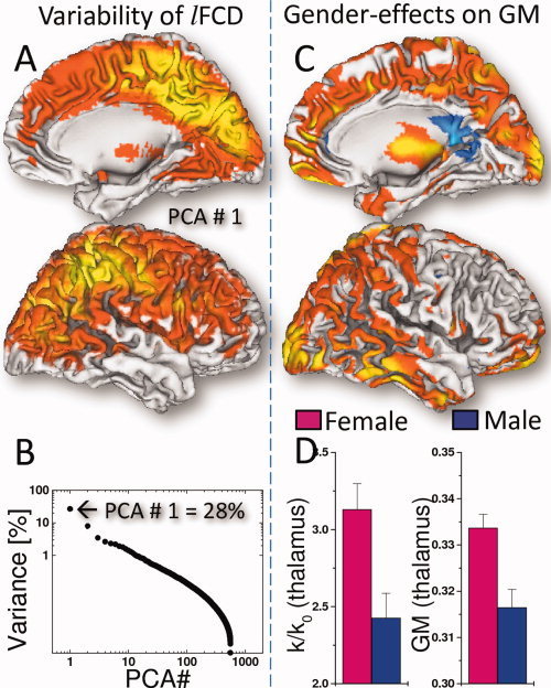
Spatial distribution of the first principal component (PCA #1) showing brain regions with high lFCD‐variance (A) and the variance of the lFCD as a function of the principal components (B). The VBM analysis revealed that women had higher gray matter (GM) density than men in occipital, parietal, temporal, and ventral prefrontal cortices and thalamus (C; red–yellow: 10−3 < P < 10−9, one‐way ANOVA). GM differences paralleled lFCD differences in the thalamus (D). Sample: 336 women and 225 men, age: 21.5 ± 2.3 years.