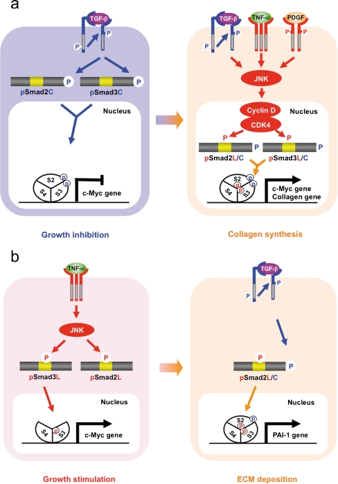
Fig. 4.
Spatial and temporal dynamics of R-Smad phosphoisoforms differing between HSC and MFB. a Phospho-Smad signaling in HSC: involvement of pSmad2L/C and pSmad3L/C pathways. TGF-β inhibits HSC growth by down-regulating c-Myc oncoprotein via the pSmad2C and pSmad3C pathways (left); TGF-β, TNF-α, and PDGF, in turn, enhance HSC growth and collagen synthesis by up-regulating the transcription of c-Myc and collagen via the pSmad2L/C and pSmad3L/C pathways (right). b Phospho-Smad signaling in MFB: involvement of the mitogenic pSmad3L and the fibrogenic pSmad2L/C pathways. TNF-α activates JNK, which phosphorylates Smad2L and Smad3L (left). After the COOH-tail phosphorylation of cytoplasmic pSmad2L by TβRI, pSmad2L/C undergoes translocation to the nucleus, where it interacts with pSmad3L and Smad4. The Smad complex then stimulates plasminogen activator inhibitor-1 (PAI-1) transcription and ECM deposition (right)

