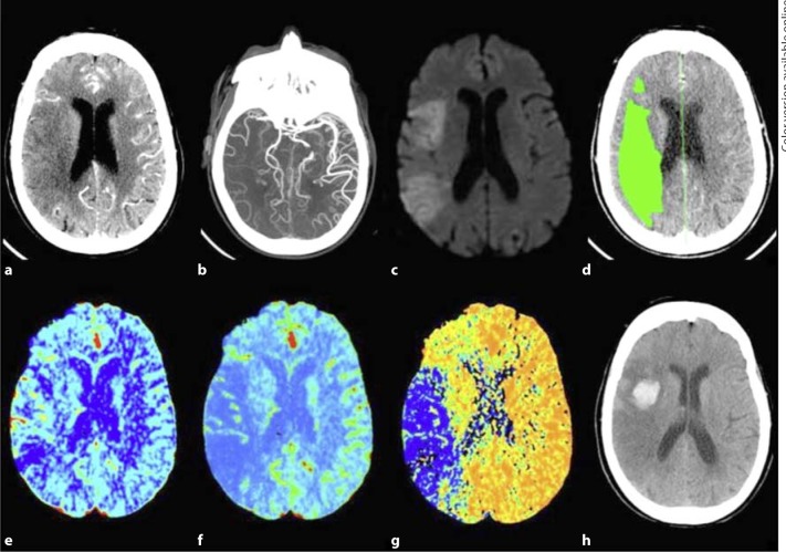
Fig. 1.
An 81-year-old female presenting 6 h after the onset of left-sided weakness and right gaze preference; not a candidate for endovascular therapy: a 5-mm thick CTA source image shows poor tissue opacification of the right MCA territory, b CTA maximum intensity projection image shows proximal right MCA occlusion with poor collateralization, c DWI shows right MCA territory infarct core, d thresholded MTT lesion (rMTT >1.3), with 140 ml total volume, e CBV map shows relative hyperemia (increased blood volume) of the cortical right MCA territory, f CBF map show decreased flow of the right MCA territory, corresponding to the DWI lesion, g MTT map shows corresponding area of prolonged transit time in the right MCA territory, mean rMTT = 4.3, and h 24 h follow-up NCCT shows HT at the anterior right MCA territory.

