
Figure 1.
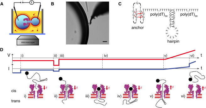
(A) Schematic depiction of the experimental setup. A single α-hemolysin pore resides in a lipid bilayer formed between two electrically contacted aqueous droplets within a lipid-oil bath. (B) The two droplets observed through a microscope (scalebar: 50 μm). (C) The hairpin of interest is connected to a stable anchor via a poly(T) sequence and extended with a second poly(T) tail on the other side acting as a capture sequence. (D) Voltage protocol used for the experiments. (i) First, a constant voltage is applied for several hundreds of milliseconds in which DNA is captured in most of the cases. (ii and iii) Then a high voltage pulse is applied to open the hairpin, regardless of whether the hairpin was already opened before. (iv) The DNA is then held in the pore at low voltage to allow reformation of the hairpin on the trans side. Finally, a voltage ramp is started (v) and the unzipping of the hairpin is observed as a sudden jump in pore current (vi).