
Figure 3.
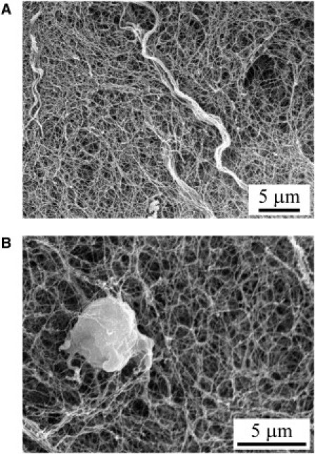
Scanning electron micrographs of the mesoglea of juvenile jellyfish in the middle of the mesoglea, ∼100–400 μm above the endoderm. (A) Thick fibers emerging from the three-dimensional network of fine fibrils. The thick fibers are woven together by many fibrils of the three-dimensional network. The fibrils are randomly and heterogeneously distributed and the size of the fibrous mesh is very variable. A similar fibrous organization was observed on pieces of mesoglea cut from adult jellyfish. (B) A mesogleal cell embedded in the fine network of fibrils.