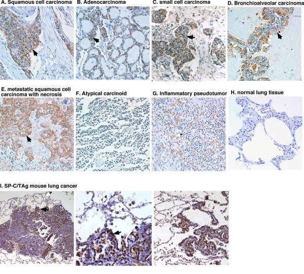
Figure 3.
IHC analysis of sPLA2-IIa expression in lung cancer specimens. Brown staining indicates positivity for sPLA2-IIa. a-e. All primary and metastatic tumors indicate positive staining for sPLA2-IIa (solid arrow). g. Some endothelial cells in new blood vessels (open arrow) and macrophages show positive staining for sPLA2-IIa in inflammatory pseudo tumor. Atypical carcinoid (f) and normal lung (h) tissue are negative staining for sPLA2-IIa. (i) sPLA2-IIa overexpression was found in the spontaneous mouse lung cancer specimens of SP-C/TAg transgenic mice (solid arrow), but not in the adjacent normal type I and II epithelial cells (open arrow)

