
Fig. 2.
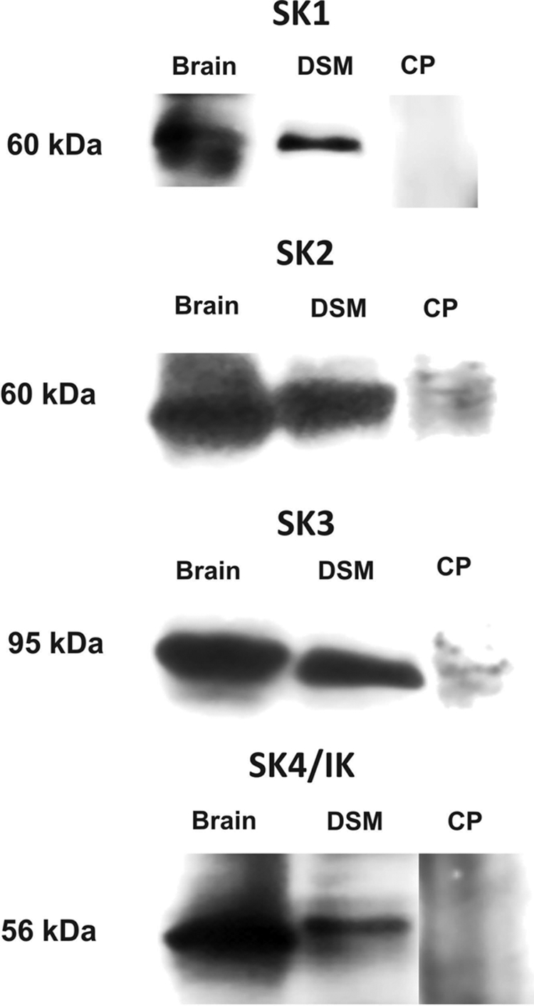
Western blot detection of SK1 (∼60 kDa), SK2 (∼60 kDa), SK3 (∼95 kDa), and IK (SK4) (∼56 kDa) channel protein expression in DSM tissues. Illustrated images are representation of at least five independent Western blot experiments based on protein extracted from five animals. Guinea pig brain was used as a positive control. The lack of immunoreactive bands in the presence of competing peptide (CP) confirmed the specificity of the primary antibodies.