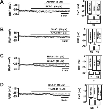
Fig. 3.
SKA-31 hyperpolarizes the resting membrane potential in freshly isolated guinea pig DSM cells. A, representative recording illustrating that SKA-31 (10 μM) hyperpolarized the resting membrane potential (RMP) in a guinea pig DSM cell. Apamin (1 μM) reversed the hyperpolarizing effect of SKA-31 (10 μM) on the resting membrane potential. Bar graph shows the effect of SKA-31 (10 μM) on the resting membrane potential in the absence and after application of 1 μM apamin (n = 5; N = 4; *, P < 0.05). B, representative recording illustrating that apamin (1 μM) pretreatment for 10 min prevented the SKA-31 (10 μM)-induced hyperpolarizing effect on the resting membrane potential in a guinea pig DSM cell. Bar graph shows the effect of apamin (1 μM) and the lack of SKA-31 (10 μM) effect in the presence of apamin on the resting membrane potential (n = 8; N = 5; P > 0.05). C, representative recording illustrating that TRAM-34 (1 μM) did not reverse the hyperpolarizing effect of SKA-31 (10 μM) on the resting membrane potential. Bar graph shows the effect of SKA-31 (10 μM) on the resting membrane potential in the absence and after application of TRAM-34 (1 μM) (n = 6; N = 5; *, P < 0.05). D, representative recording showing that pretreatment of DSM cells with TRAM-34 (1 μM) for 10 min did not change the SKA-31 (10 μM)-induced hyperpolarizing effect on the resting membrane potential in guinea pig DSM cells. Bar graph shows the lack of TRAM-34 (1 μM) effect alone and the hyperpolarizing effect of SKA-31 (10 μM) in the presence of TRAM-34 (1 μM) (n = 11; N = 6; *, P < 0.05). All experiments were performed in the presence of paxilline (100 nM). NS, nonsignificant.

