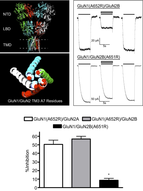
Fig. 1.
Ethanol inhibition of A7 arginine-substituted NMDA receptors. A, top panel shows a model of the GluN1/GluN2A receptor with extracellular amino terminal domain (ATD), LBD, and transmembrane domain (TMD) (dashed line represents approximate location of the plasma membrane). Bottom panel shows a top-down view of TM3 helices (GluN1, white/green; GluN2A, blue/red) with alanines at position 7 of the conserved SYTANLAAF domain shown as space-filling molecules. B, traces show representative currents from single cells expressing A7R mutants [top, GluN1(A652R)/GluN2B; bottom, GluN1/GluN2B(A651R)]. In each set of traces, currents were activated by switching from a solution containing magnesium to one containing calcium (both at 10 mM; solid line) in the absence and presence of 100 mM ethanol (▩). C, summary of effects of 100 mM ethanol on spontaneous currents from arginine-substituted A7 mutants. Dashed lines show mean ethanol inhibition of GluN1/GluN2A (41.7 ± 4.9%; blue line) and GluN1/GluN2B (49.4 ± 4.7%; red line) for comparison. Data are means ± S.E.M. from 8 to 14 cells for each receptor combination. *, value significantly different from that for GluN1(A652R)/GluN2B; unpaired t test (p < 0.0001).

