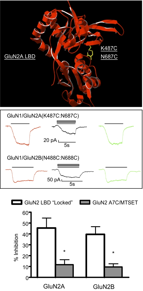
Fig. 4.
Ethanol inhibition of NMDA receptors with locked ligand-binding domains. A, model of the LBD of the GluN2A subunit showing cysteines substituted for lysine at position 487 (K487C) and asparagine at position 687 (N687C). The proximity of these cysteine-substituted resides locks the LBD in the closed (activated) conformation. B, traces show representative currents from single cells expressing locked LBD mutants [top, GluN1/GluN2A(K487C:N687C); bottom, GluN1/GluN2B(N488C:N688C)]. Currents were activated by switching from a recording solution containing 10 mM MgCl2 to one containing 10 mM CaCl2 (solid line, red and green traces) alone or with 100 mM ethanol (▩, black trace). C, summary of effects of 100 mM ethanol on spontaneous currents from locked LBD mutants (□). Ethanol inhibition of MTSET-treated A7C mutants is shown for comparison (▩; taken from Fig. 3). Data are the mean ± S.E.M. from 7 to 18 cells for each receptor combination. *, value significantly different from that for the corresponding locked LBD GluN2 mutant; unpaired t test (p < 0.01).

