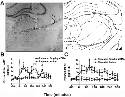
Fig. 1.
A, coronal section of the rat brain illustrating electrode placement in the VPM thalamus. Low-power photomicrograph demonstrating the tip of the microelectrode bundle in the VPM thalamus. The section was stained with neutral red. B, effects of repeated systemic administration of MDMA on extracellular 5-HT levels in the rat VPM thalamus. Serotonin levels were measured using in vivo microdialysis with HPLC. Significant prolonged fluctuating increases in extracellular 5-HT were elicited by a challenge injection of MDMA after repeated MDMA administration (n = 6 animals). Repeated administration of saline (n = 4 animals) did not elicit any significant changes in extracellular 5-HT. C, effects of repeated systemic administration of MDMA on extracellular NE levels in the rat VPM thalamus. Norepinephrine levels in the rat VPM thalamus were measured using in vivo microdialysis with HPLC. Significant increases in extracellular NE were elicited by a challenge injection of MDMA after repeated MDMA administration (n = 6 animals). Repeated administration of saline (n = 4 animals) did not elicit any significant changes in extracellular NE. All data are presented as the mean ± S.E.M.; *, significant changes (Dunnett's post hoc test, p > 0.05).

