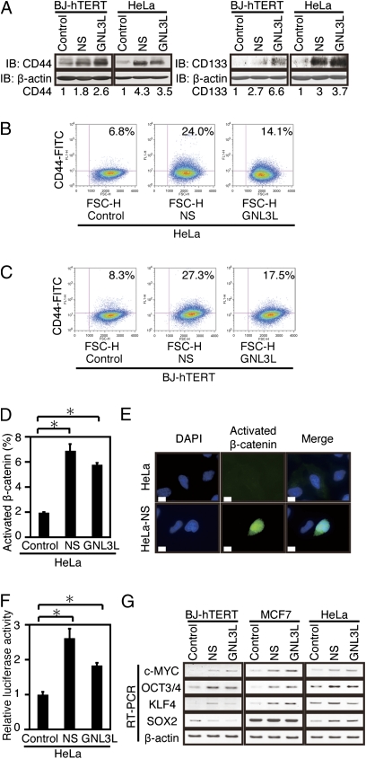
Fig. 3.
Effects of NS or GNL3L on cancer stem cell markers. Effects of overexpressing NS or GNL3L on CD44 protein expression as assessed by immunoblotting (A) or flow cytometry (B and C). Cells were stained with FITC-conjugated anti-CD44 (Leu-44) antibody. IB, immunoblot. (B) Fractions of HeLa cells expressing high levels of CD44 were 6.8% (control vector), 24.0% (NS), and 14.1% (GNL3L). (C) Fractions of BJ-hTERT cells expressing high levels of CD44 were 8.3% (control vector), 27.3% (NS), and 17.5% (GNL3L). (D) Percentage of cells that harbor nuclear activated β-catenin in cells expressing a control vector, NS, or GNL3L is shown. *P < 0.05. (E) Effects of overexpressing NS or GNL3L on β-catenin function. Representative immunofluorescence images are shown. HeLa cells (Upper) and HeLa cells expressing NS (Lower) were stained with an antibody that recognizes unphosphorylated (active) β-catenin and visualized with Alexa Fluor 488 (Invitrogen)-conjugated anti-mouse IgG (green, magnification: 600×); DAPI (blue) indicates DNA. (Scale bar = 10 μm.) (F) TOPFLASH-Luc luciferase reporter activity. A renilla luciferase expression plasmid, pRL-SV40, was used as an internal control for transfection efficiency. *P < 0.05. (G) Effects of overexpressing NS or GNL3L on the expression of c-MYC, OCT3/4, KLF4, and SOX2.

